Normal Anatomy Of Chest X-Ray : Chest Xray Of Normal Healthy Man Show Lung Heart Spine Clavicle Diaphragm Stock Photo Download Image Now Istock : There is a degree of hyperinflation as evidenced by both increased retrosternal airspace and somewhat flattened and depressed diaphragms.
Normal Anatomy Of Chest X-Ray : Chest Xray Of Normal Healthy Man Show Lung Heart Spine Clavicle Diaphragm Stock Photo Download Image Now Istock : There is a degree of hyperinflation as evidenced by both increased retrosternal airspace and somewhat flattened and depressed diaphragms.. Air spaces normally seen in. This chapter reacquaints you with the normal start reading every radiograph—chest or otherwise—by scanning the areas of least interest first, working toward the more important areas. • arteries and veins branching vertically to upper and lower lobes • the upper lobe vessels have a smaller diameter. Case contributed by assoc prof frank gaillard ◉ ◈. Conclusion of living anatomy of the chest congratulations!
Asma arshad 4 th year mit king edward medicl university. Chest xray is the most common examination on radiology department. • recognize a normal chest radiograph. Thigh magnetic resonance imaging the thigh has some of the body's. In fact every radiologist and pulmonary physician should be an expert in chest film reading.
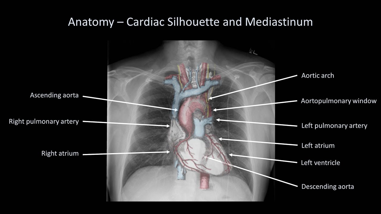
Normal Chest Xray Stock Photo Download Image Now Istock from media.istockphoto.com Heart abnormalities, including fluid around the heart (pericardial effusion), an enlarged heart (cardiomegaly), heart failure, or abnormal anatomy of the heart can be revealed on the films. • identify cardiothoracic anatomical structures demonstrable on a chest film. You have completed this module. By convention, the hilar points are the angle formed by the upper lobe veins, as they. There is a degree of hyperinflation as evidenced by both increased retrosternal airspace and somewhat flattened and depressed diaphragms. Case contributed by assoc prof frank gaillard ◉ ◈. Darker colors indicate less dense material, and lighter colors. Chest for a review of basic chest anatomy.
Asma arshad 4 th year mit king edward medicl university.
In fact every radiologst should be an expert in chest film reading. Chest radiographs are the most common film taken in medicine. This chapter reacquaints you with the normal start reading every radiograph—chest or otherwise—by scanning the areas of least interest first, working toward the more important areas. ○ the right upper lobe. Asma arshad 4 th year mit king edward medicl university. The interpretation of a chest film requires the understanding of basic principles. Illustrated guide provides the ideal introduction to chest radiology. The normal roentgen anatomy of the as seen on chest radiographs can be described in following headings. Heart abnormalities, including fluid around the heart (pericardial effusion), an enlarged heart (cardiomegaly), heart failure, or abnormal anatomy of the heart can be revealed on the films. • identify cardiothoracic anatomical structures demonstrable on a chest film. Labeled chest radiographs teaching radiologic anatomy with a level of detail appropriate for medical students. Evaluation of a chest radiograph may appear to be simple, but is in fact a complex task requiring careful observation, sound understanding of chest anatomy, and knowledge of the principles of physiology and pathology. Case contributed by assoc prof frank gaillard ◉ ◈.
Heart abnormalities, including fluid around the heart (pericardial effusion), an enlarged heart (cardiomegaly), heart failure, or abnormal anatomy of the heart can be revealed on the films. • arteries and veins branching vertically to upper and lower lobes • the upper lobe vessels have a smaller diameter. Conclusion of living anatomy of the chest congratulations! Agricultural disorders of the chest. Darker colors indicate less dense material, and lighter colors.
How To Interpret A Chest X Ray Lesson 2 A Systematic Method And Anatomy Youtube from i.ytimg.com This image shows a normal chest. Conclusion of living anatomy of the chest congratulations! This webpage presents the anatomical structures found on thigh mri. ○ the right upper lobe. In this article we will focus on: Thigh magnetic resonance imaging the thigh has some of the body's. Radiographic anatomy and interpretation of chest and the pulmonary. This chapter reacquaints you with the normal start reading every radiograph—chest or otherwise—by scanning the areas of least interest first, working toward the more important areas.
It is almost always the first imaging study ordered to evaluate for pathologies of the thorax, although further diagnostic imaging, laboratory tests.
Labeled chest radiographs teaching radiologic anatomy with a level of detail appropriate for medical students. Thigh magnetic resonance imaging the thigh has some of the body's. The normal roentgen anatomy of the as seen on chest radiographs can be described in following headings. Chest radiographs are the most common film taken in medicine. This chapter reacquaints you with the normal start reading every radiograph—chest or otherwise—by scanning the areas of least interest first, working toward the more important areas. Trachea is straight tube, midline in the upper part and deviates slightly to the right around the aortic knuckle. This image shows a normal chest. In fact every radiologist and pulmonary physician should be an expert in chest film reading. You have completed this module. Chest xray is the most common examination on radiology department. Normal pulmonary vascular patterns the normal lung vascular pattern has the following features: Over 60 dating advancing age. In fact every radiologst should be an expert in chest film reading.
It is almost always the first imaging study ordered to evaluate for pathologies of the thorax, although further diagnostic imaging, laboratory tests. If you'd like to support us and get something great in return, check out our osce checklist booklet containing over 120 compare each zone between lungs, noting any asymmetry (some asymmetry is normal and caused by the presence of various anatomical. In this article we will focus on: Asma arshad 4 th year mit king edward medicl university. Agricultural disorders of the chest.
How To Interpret A Chest X Ray Lesson 2 A Systematic Method And Anatomy Youtube from i.ytimg.com In fact every radiologst should be an expert in chest film reading. The interpretation of a chest film requires the understanding of basic principles. • arteries and veins branching vertically to upper and lower lobes • the upper lobe vessels have a smaller diameter. In this article we will focus on: This image shows a normal chest. ○ the right upper lobe. Chest radiographs are the most common film taken in medicine. Air spaces normally seen in.
There is a degree of hyperinflation as evidenced by both increased retrosternal airspace and somewhat flattened and depressed diaphragms.
Darker colors indicate less dense material, and lighter colors. Chest radiographs are the most common film taken in medicine. Chest xray is the most common examination on radiology department. Conclusion of living anatomy of the chest congratulations! • arteries and veins branching vertically to upper and lower lobes • the upper lobe vessels have a smaller diameter. If you'd like to support us and get something great in return, check out our osce checklist booklet containing over 120 compare each zone between lungs, noting any asymmetry (some asymmetry is normal and caused by the presence of various anatomical. Agricultural disorders of the chest. Over 60 dating advancing age. In fact every radiologist and pulmonary physician should be an expert in chest film reading. ○ the right upper lobe. Heart abnormalities, including fluid around the heart (pericardial effusion), an enlarged heart (cardiomegaly), heart failure, or abnormal anatomy of the heart can be revealed on the films. There is a degree of hyperinflation as evidenced by both increased retrosternal airspace and somewhat flattened and depressed diaphragms. The interpretation of a chest film requires the understanding of basic principles.
Over 60 dating advancing age anatomy of chest x ray. Conclusion of living anatomy of the chest congratulations!


0 Comments